

Intelligent Automation
Reduce repetitive tasks with AI-powered reconstruction, detection and measurement, making everyday workflows faster and more efficient.
_2.png)
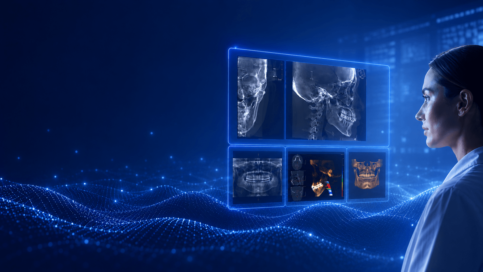
Our Intelligent Imaging Platform seamlessly integrates advanced imaging,
AI-powered analysis and clinical workflows. It transforms complex 3D data into intuitive, actionable clinical insights, helping clinicians analyze faster, make more confident decisions and dedicate more time to patient care.
Reduce repetitive tasks with AI-powered reconstruction, detection and measurement, making everyday workflows faster and more efficient.

Transform complex 3D data into intuitive, actionable clinical insights through intelligent visualization and synchronized multi-view imaging.

Everything from routine diagnostics to advanced specialty applications in one connected platform. Review images, plan implants, analyze orthodontic cases and evaluate TMJ without interrupting your workflow.

AI provides fast, reliable analysis to support clinical decisions, helping clinicians work with greater confidence and consistency.
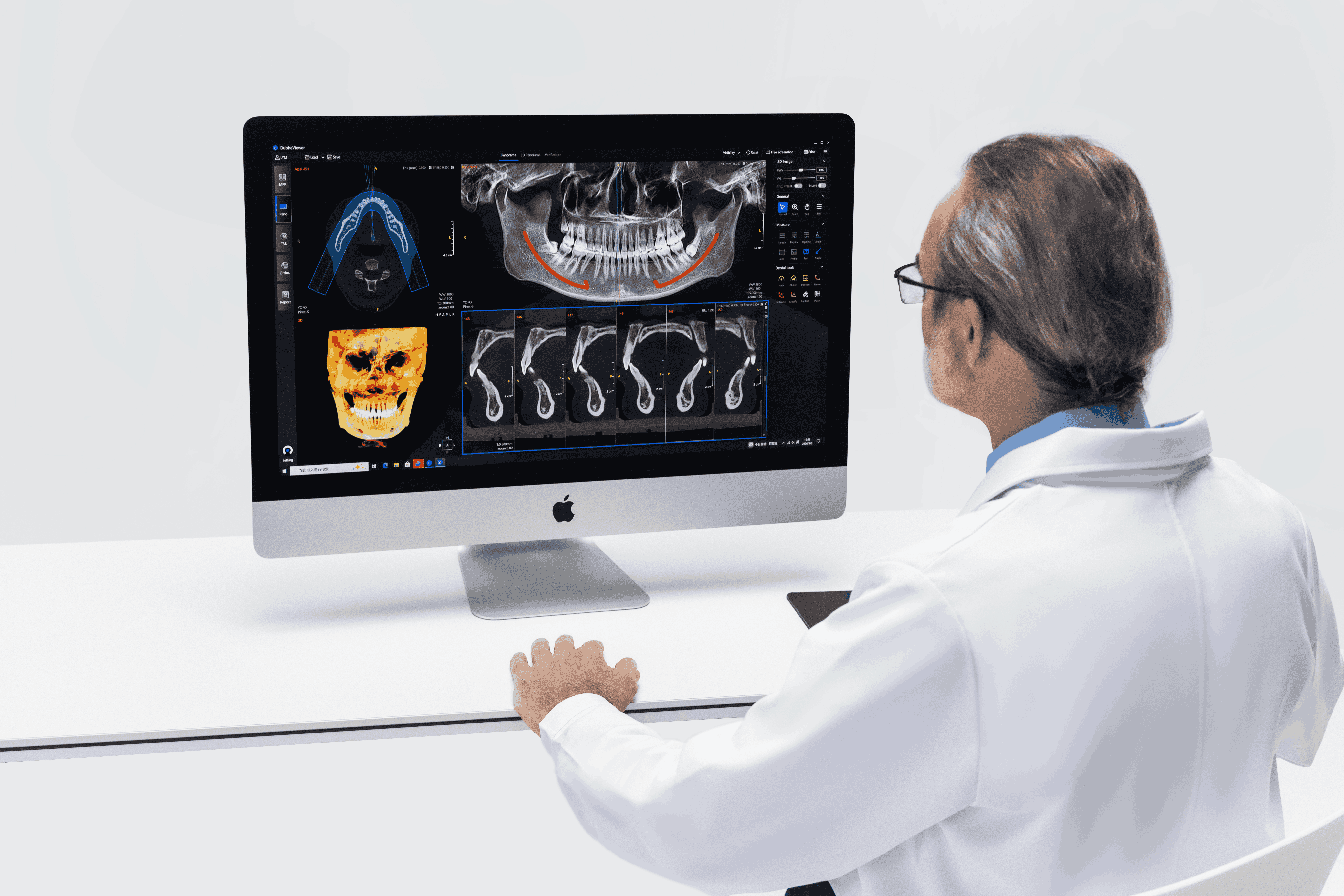
Navigate complex 3D anatomy
with an intuitive MPR workspace
designed to simplify image
interpretation and improve
clinical efficiency.
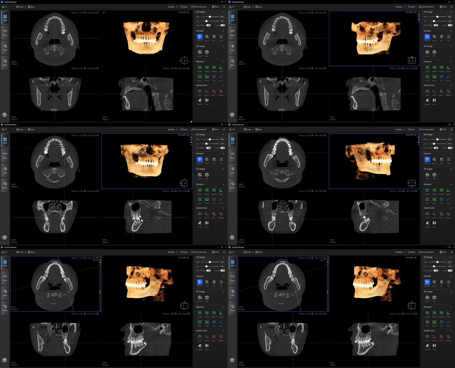
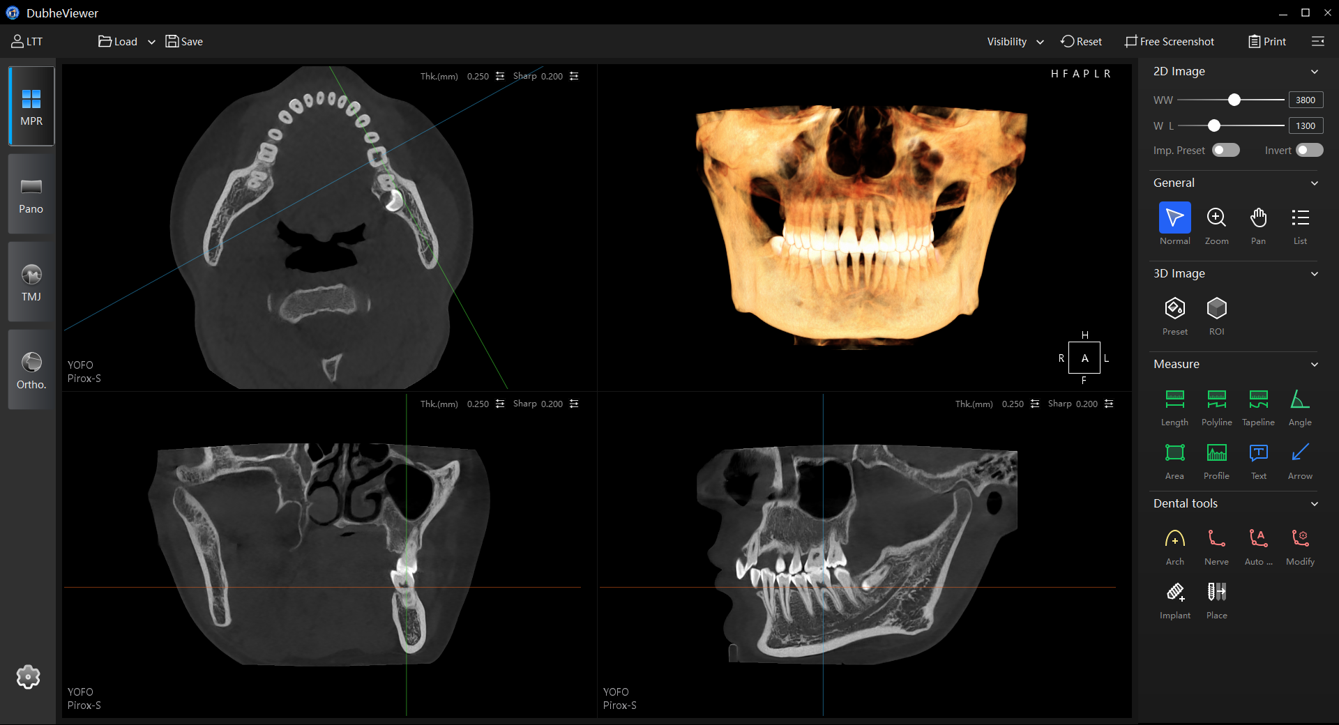
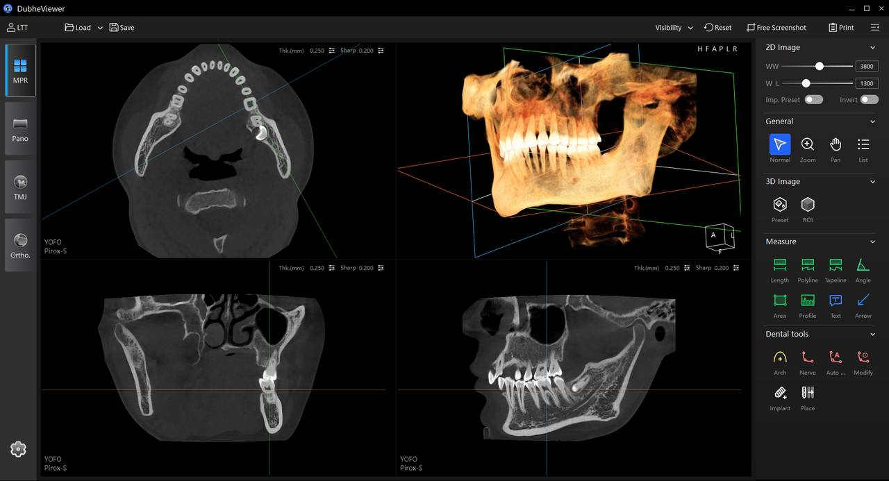
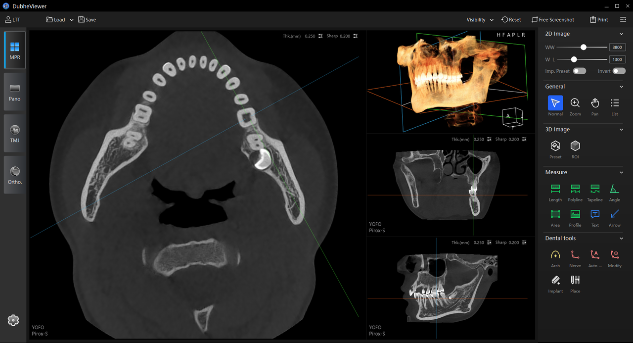
Click any point in the 3D view to instantly synchronize the coronal, sagittal and axial planes. Intuitive real-time navigation simplifies spatial orientation and helps clinicians locate anatomical structures with confidence.
_2.png)
_2.png)
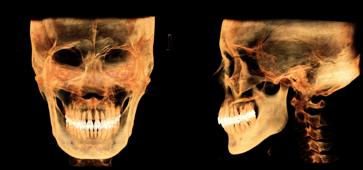
Automatically remove cervical spine structures from the 3D reconstruction to reveal a clear view of the dental arches. Improve case presentation and treatment communication for implant planning, orthodontics and other clinical applications.
_1.png)
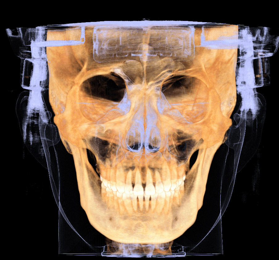
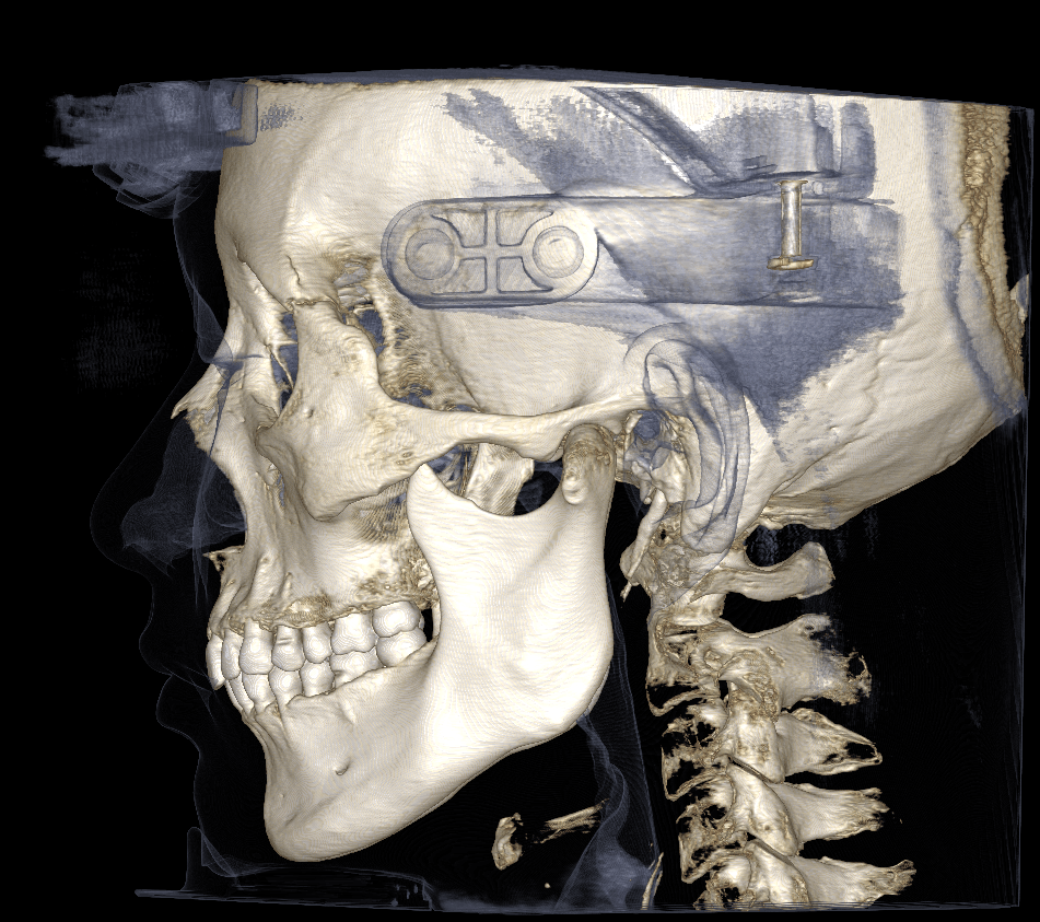
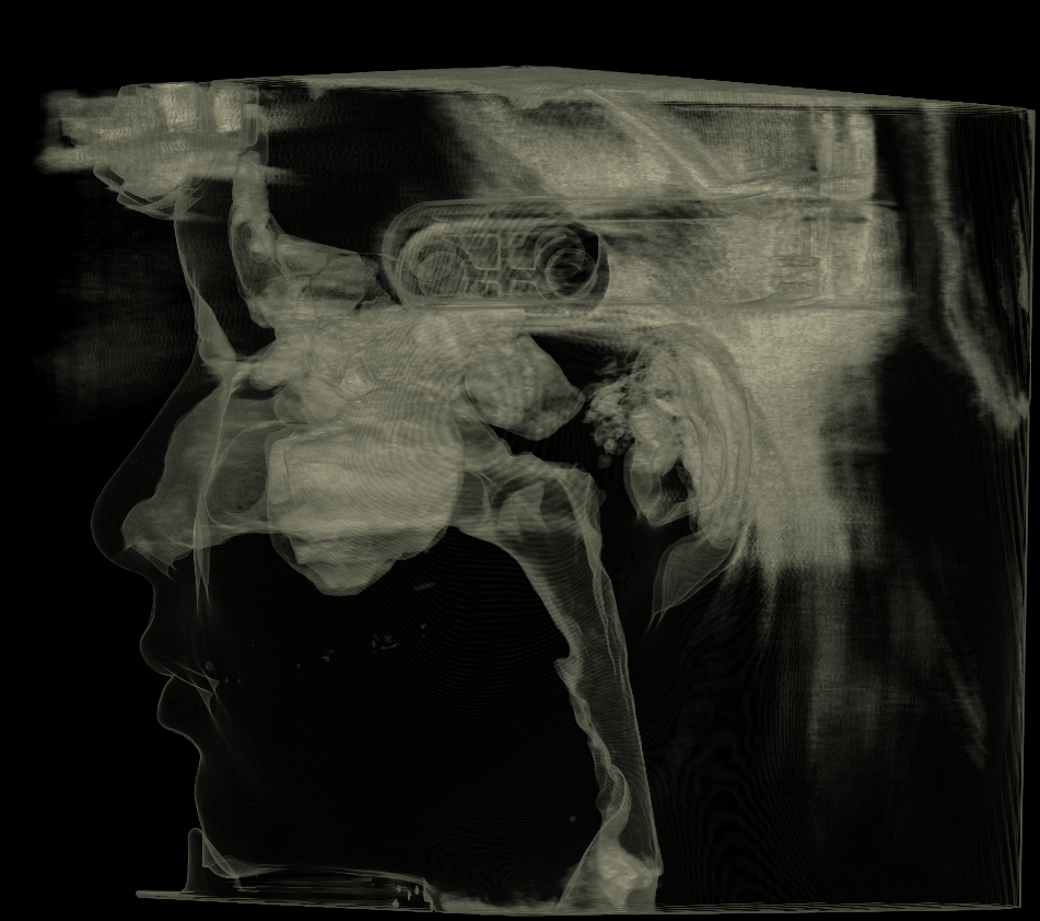
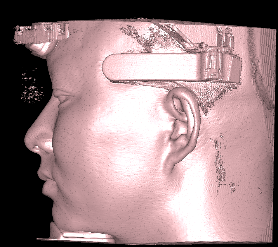
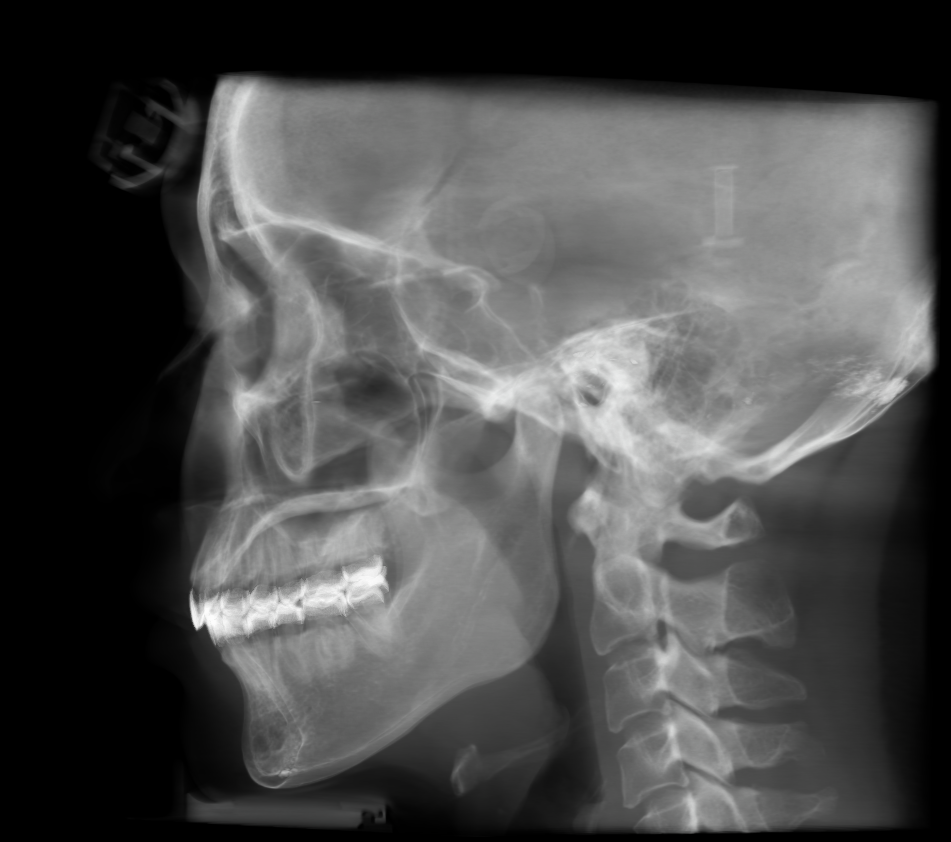
Switch instantly between seven dedicated rendering presets designed for different clinical scenarios. Highlight bone structures, dentition or other anatomical features with a single click for faster diagnosis and more effective patient communication.
Available for medium and large FOV scans
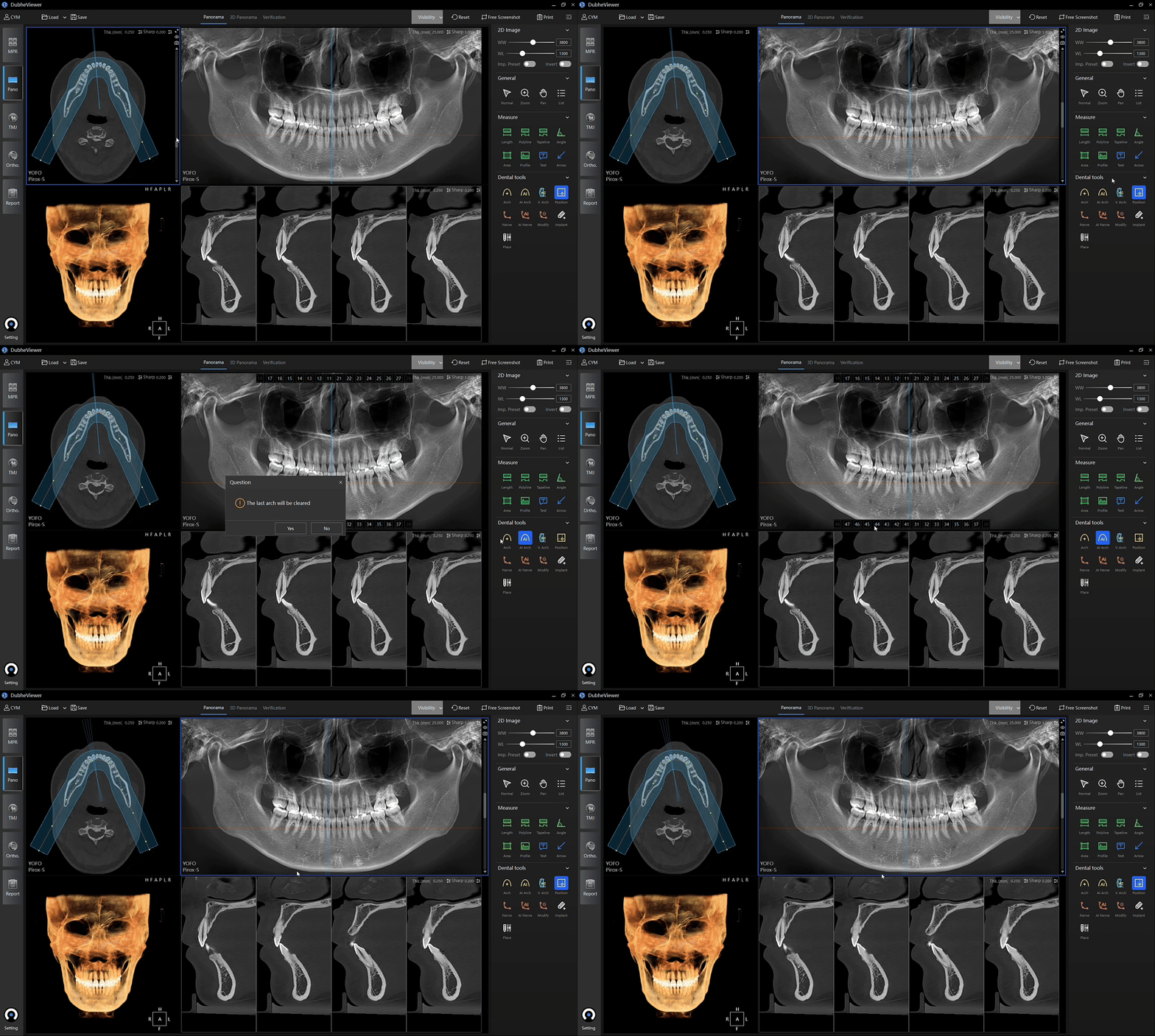
Automatically generate optimized panoramic images from CBCT data with a single click. Intelligent algorithms determine the optimal panoramic curve and layer thickness, eliminating time-consuming manual adjustments. The result is standardized, consistent image quality with fewer operating steps and a simpler everyday workflow.
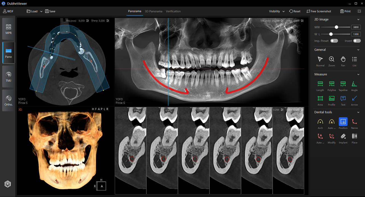
Automatically identify and trace the mandibular canal from CBCT data, eliminating time-consuming
manual tracing. Accurate canal visualization supports safer implant planning, streamlines risk
assessment and gives clinicians greater confidence throughout the planning process.

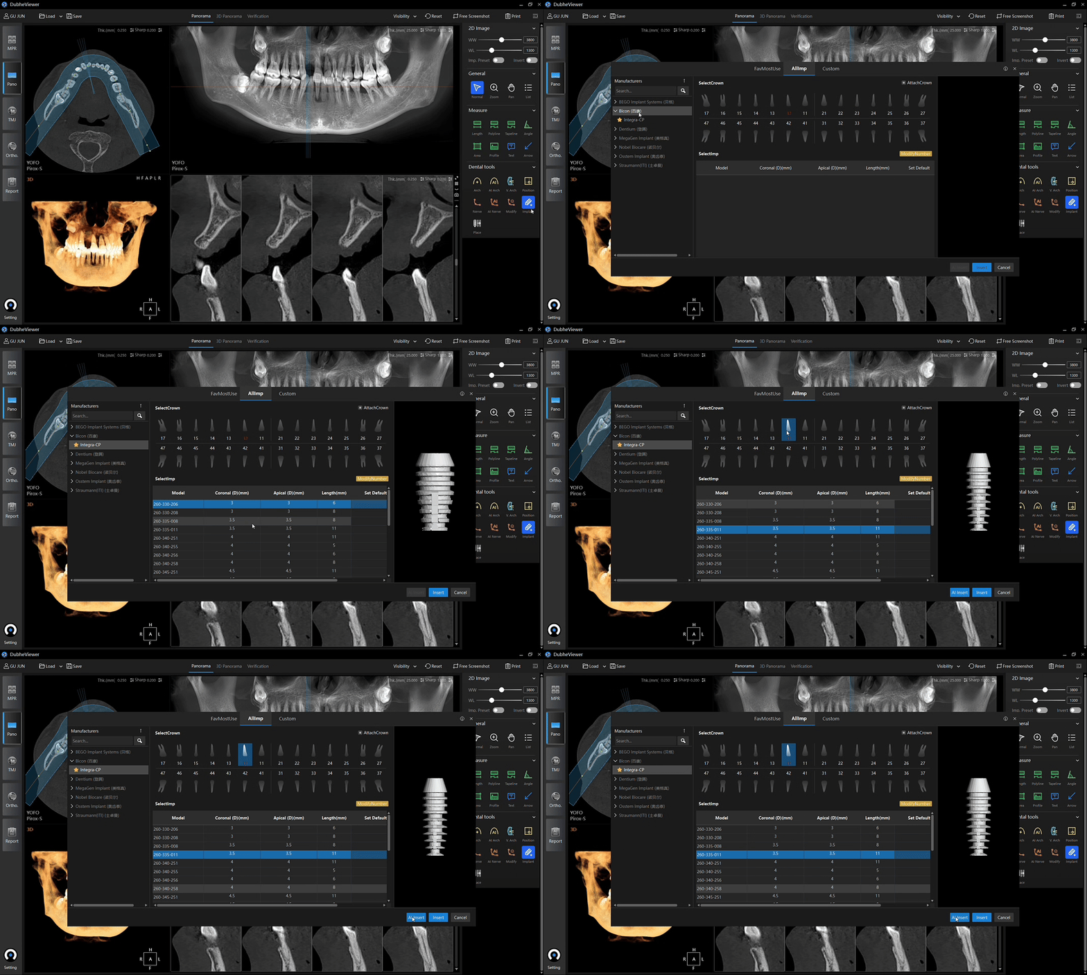
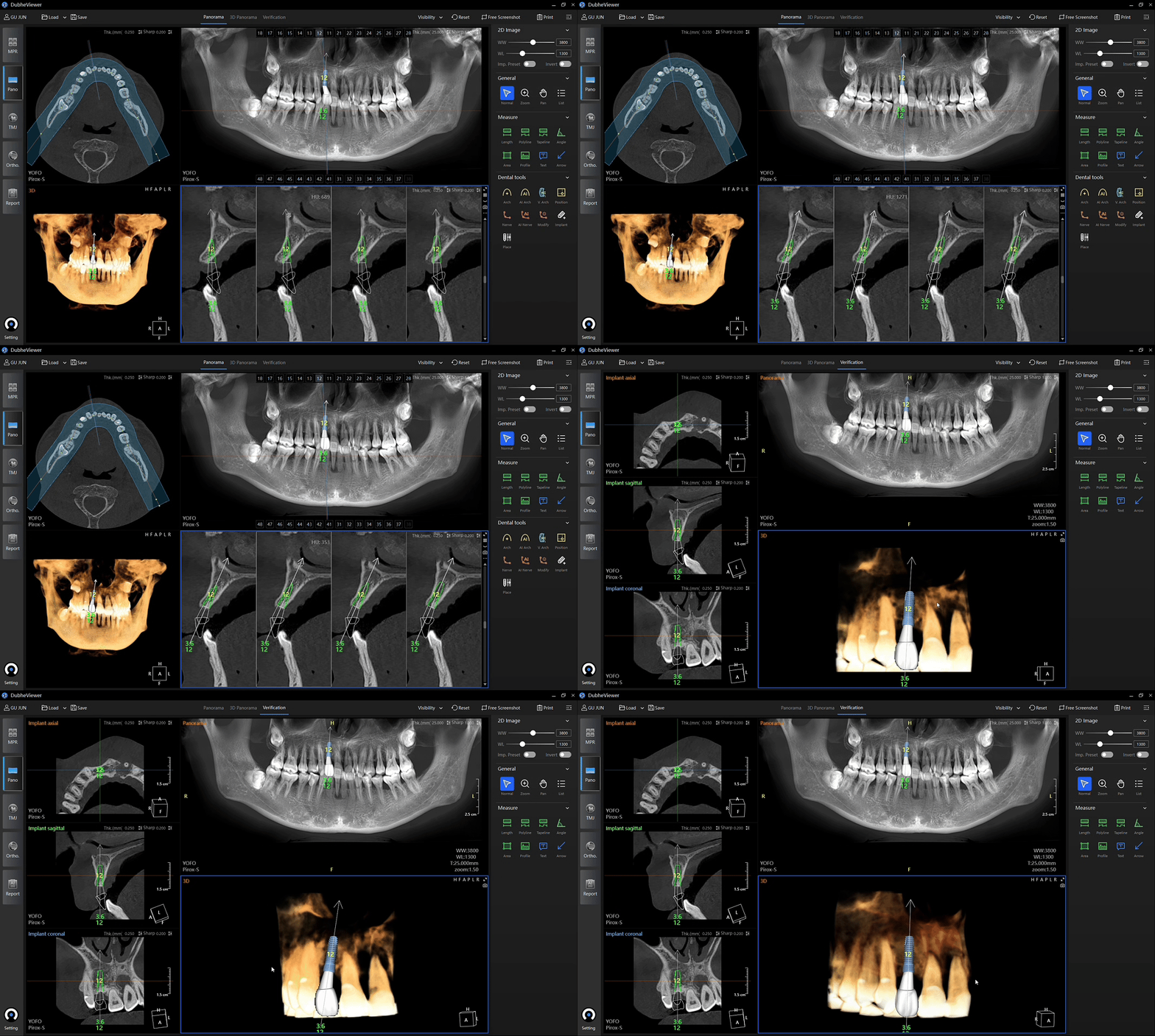
Plan implant placement directly within the panoramic or MPR view using the integrated implant library. Easily adjust implant position, angulation, depth and diameter through an intuitive workflow that simplifies preoperative planning.
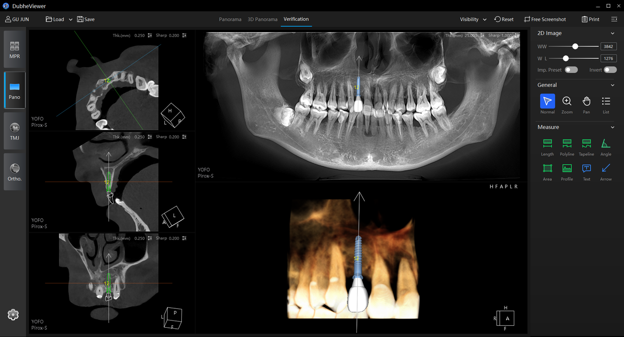
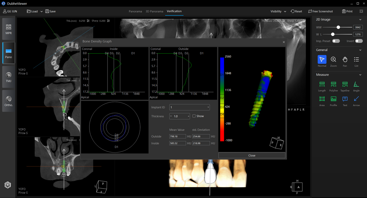
Validate implant plans with a single click by assessing implant positioning, angulation, surrounding anatomical structures and bone density. Clear visual insights help clinicians refine treatment plans with greater confidence and communicate more effectively with patients.
Available for 20 × 18 cm large FOV scans
One-click TMJ localization enables clinicians to examine condylar morphology, position and its relationship with the glenoid fossa from any angle. Synchronized 2D sectional views and 3D reconstructions provide both high-resolution anatomical detail and clear spatial context, supporting a more comprehensive assessment of temporomandibular joint disorders.
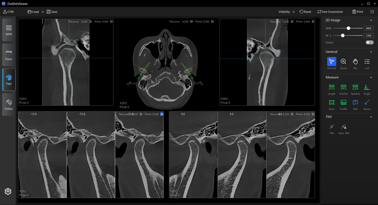
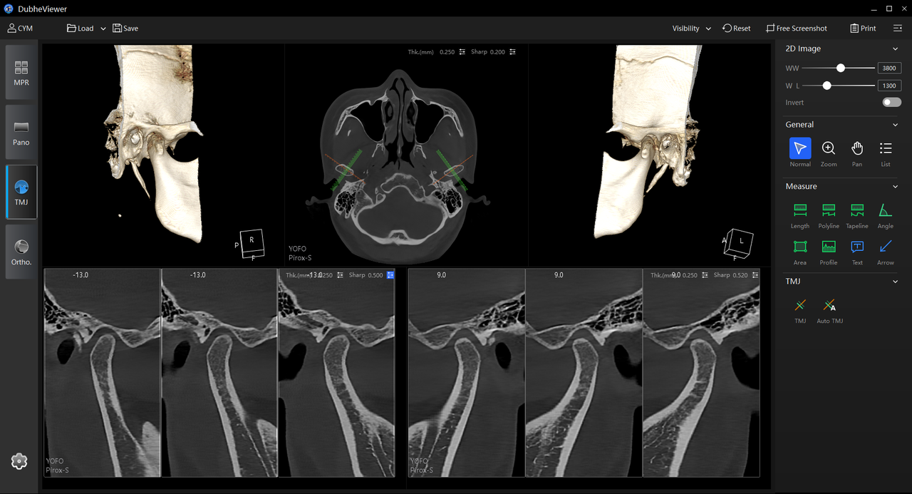
Not available for images acquired in 8 × 8 cm or 4 × 4 cm modes
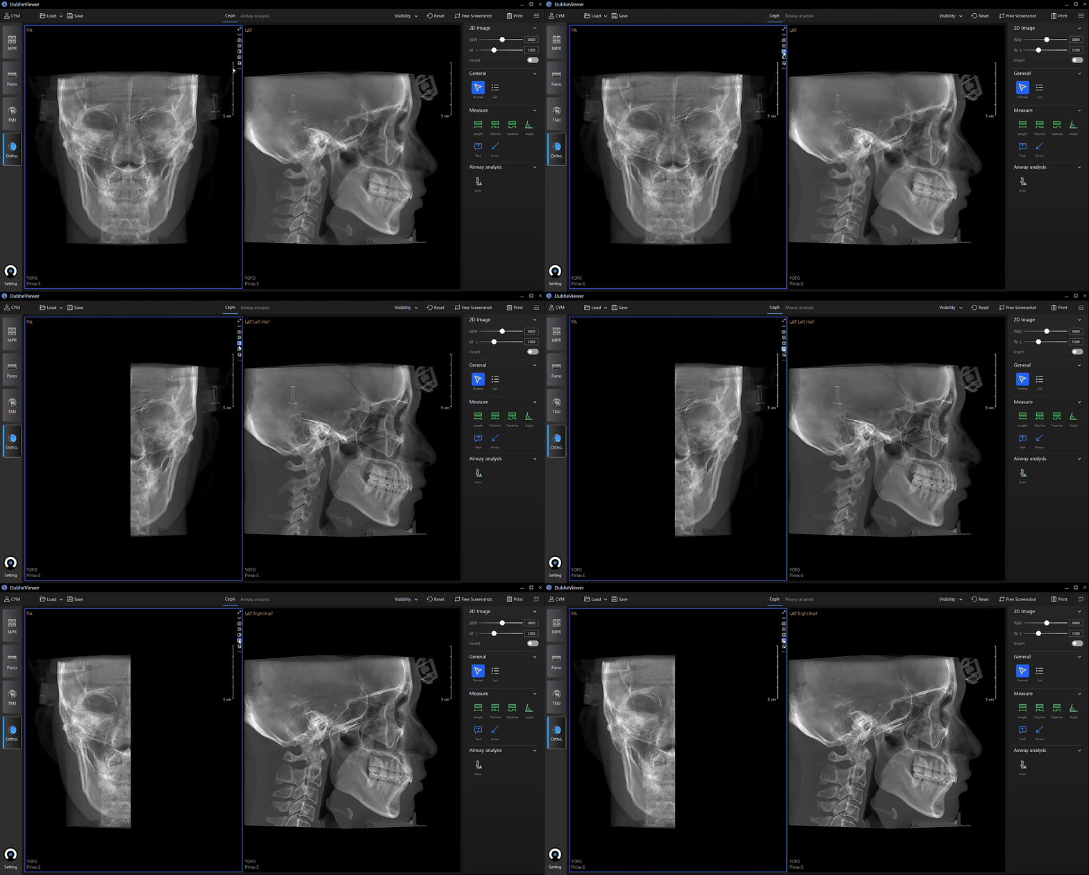
Advanced intelligent tools elevate conventional cephalometric analysis, delivering greater precision, efficiency and confidence for orthodontic and orthognathic treatment planning.
Available in 20 × 18 cm mode
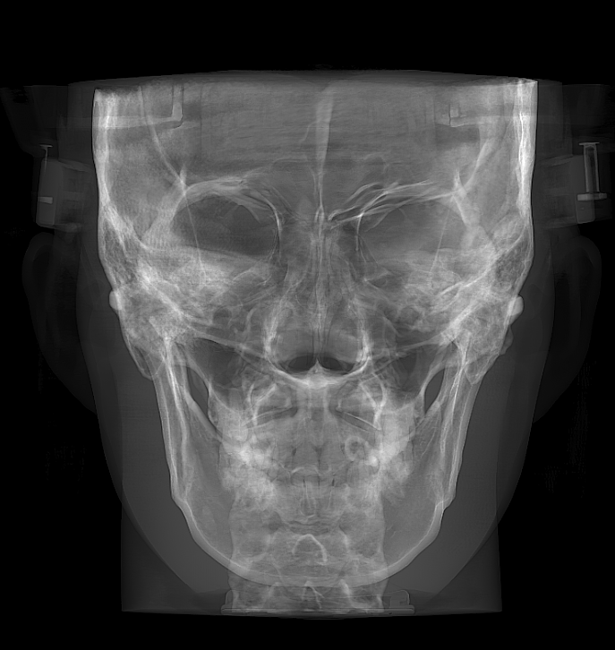
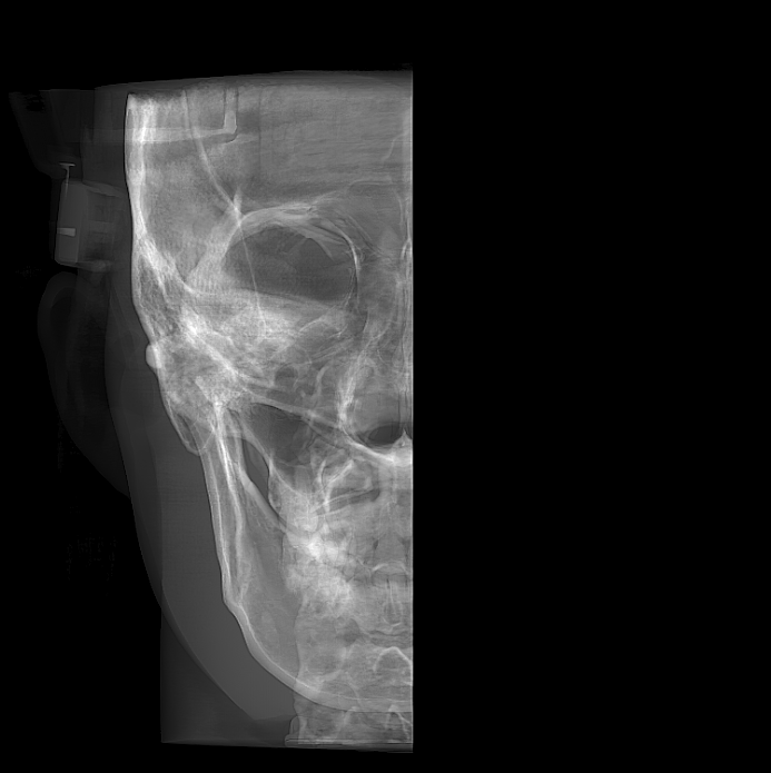
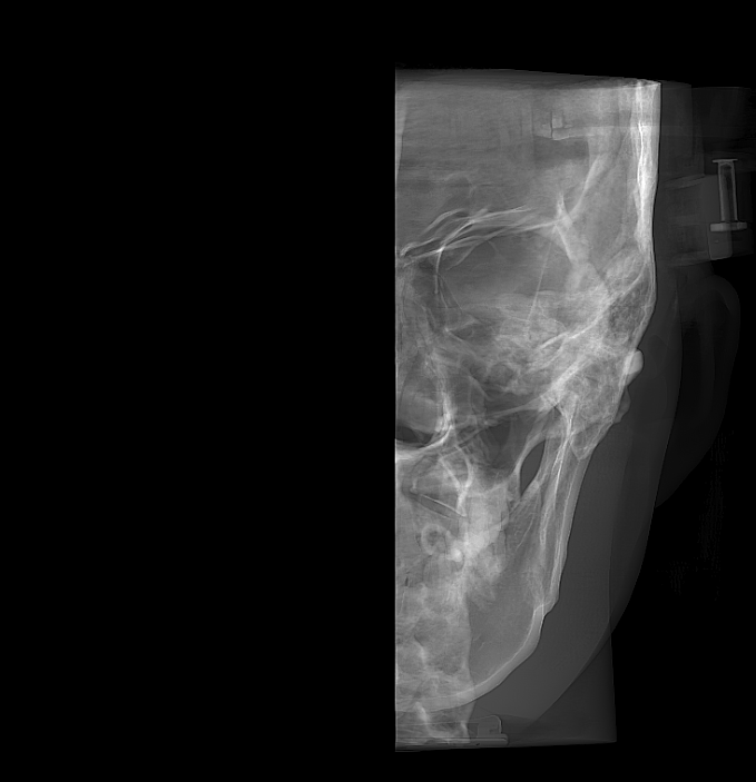
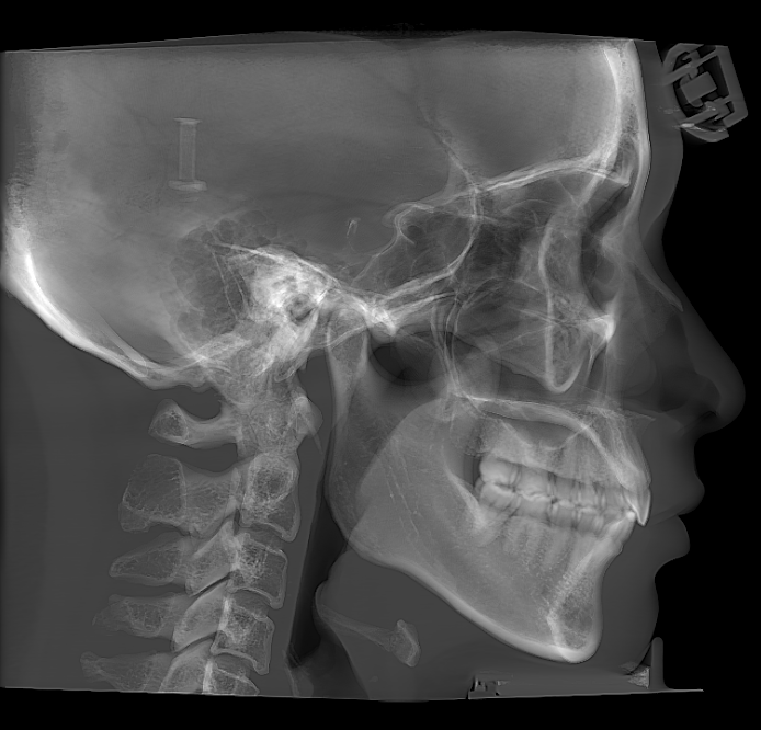
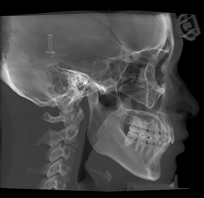
Beyond standard frontal and lateral cephalometric views, unilateral analysis allows independent adjustment and evaluation of the left and right sides using crosshair reference lines. Separate measurement of each side provides greater precision for patients with facial asymmetry, supporting more confident orthodontic and orthognathic treatment planning.
Enabled by default in 20 × 18 cm mode and available as an optional setting in 17 × 10.5 cm mode
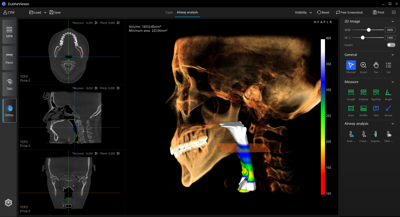
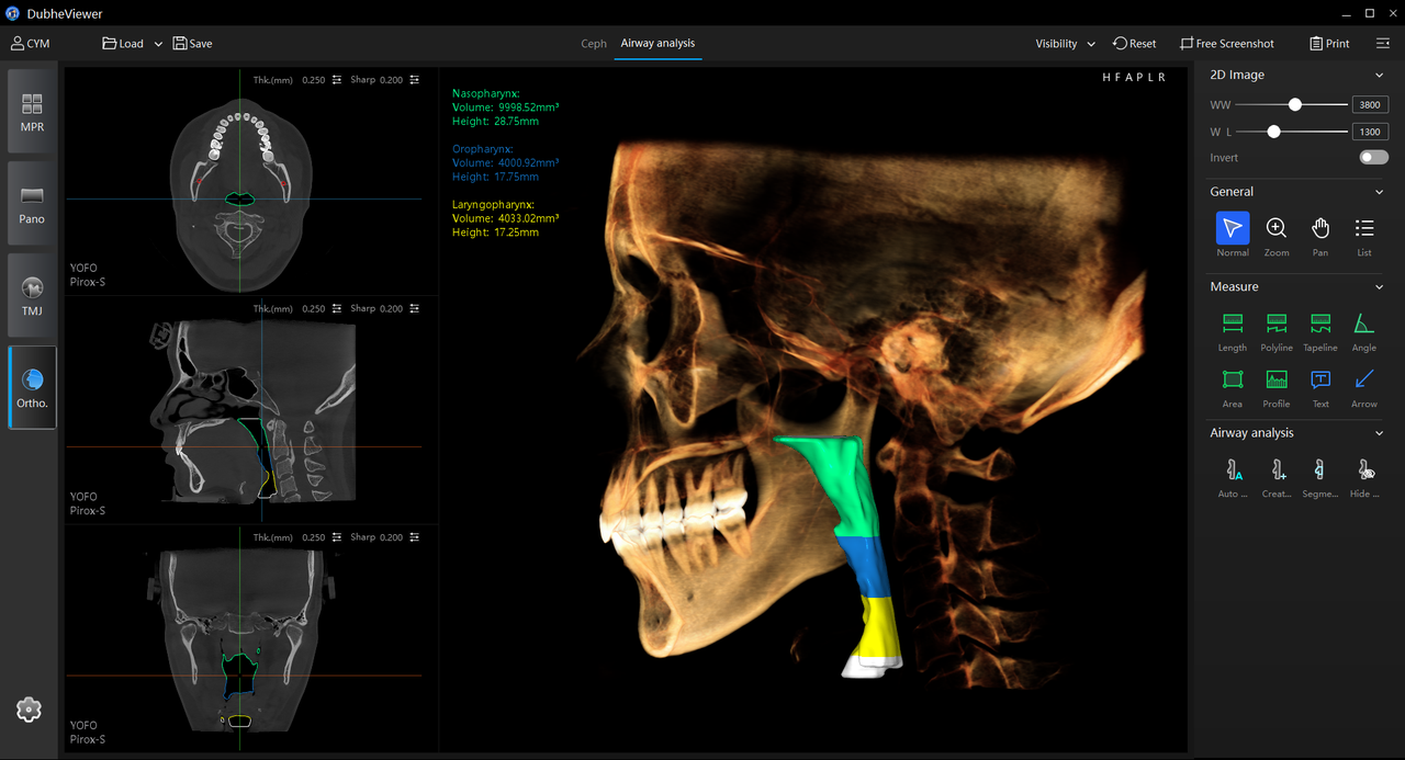
Automatically identify and segment the upper airway, including the nasopharyngeal, retropalatal and retroglossal regions, with a single click. The software locates the narrowest airway region and calculates cross-sectional areas across individual segments, providing quantitative insights for airway assessment and the screening of obstructive sleep apnea (OSA).
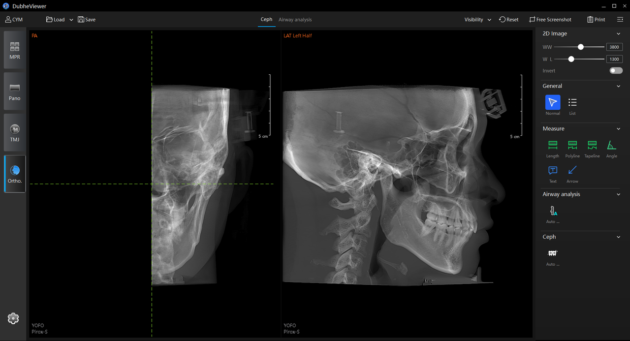
An integrated orthodontic workspace that combines intelligent automation, treatment analysis and professional reporting to streamline every stage of the clinical workflow.
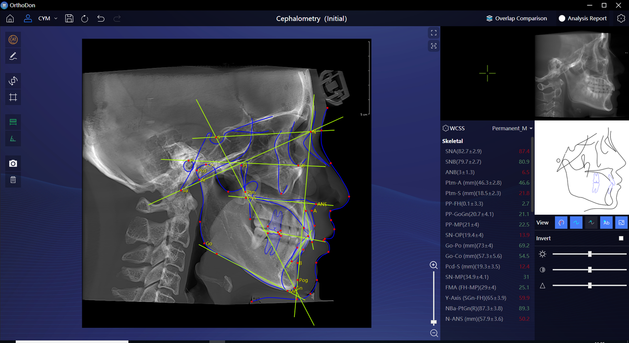
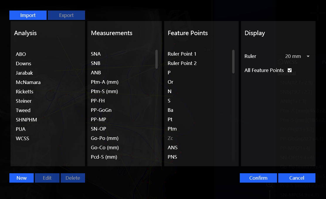
Automatically identify hundreds of cephalometric landmarks and generate measurements using established analyses such as Steiner, Tweed and WCSS. Clinicians can simply review and fine-tune results, dramatically reducing repetitive work while accelerating treatment planning.
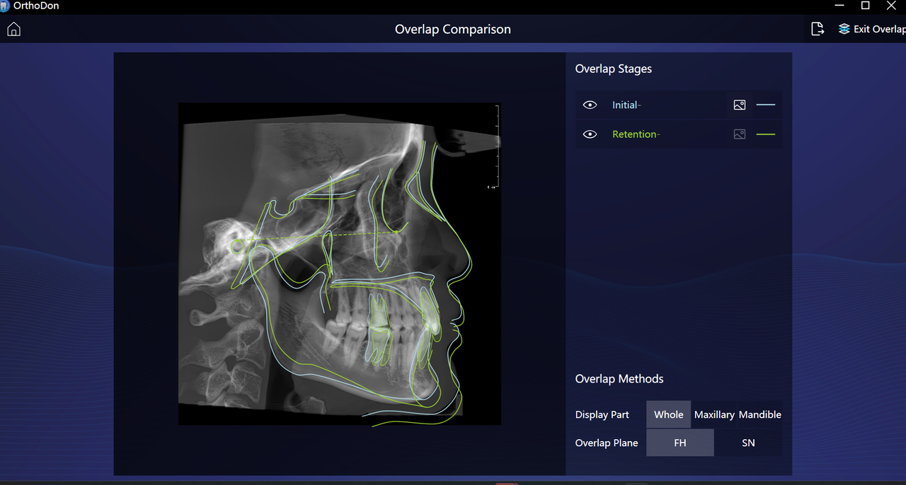
Compare cephalometric images from different treatment stages through precise superimposition. Visualize tooth movement and skeletal changes over time, making treatment outcomes easier to evaluate and explain to patients.
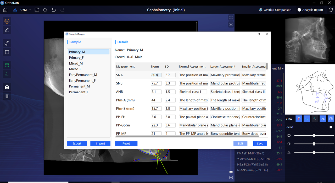
Customize reference values for any cephalometric analysis to match individual clinical preferences or patient populations. Flexible standards enable more personalized evaluation while adapting to different diagnostic philosophies.
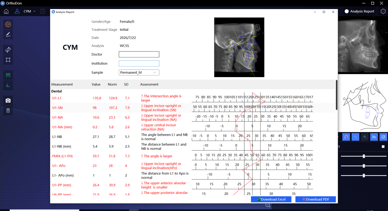
Generate professional orthodontic reports with a single click, including measurements, highlighted findings and comparison charts. Export reports as PDF or image files for documentation, printing, patient communication or multidisciplinary case consultation.
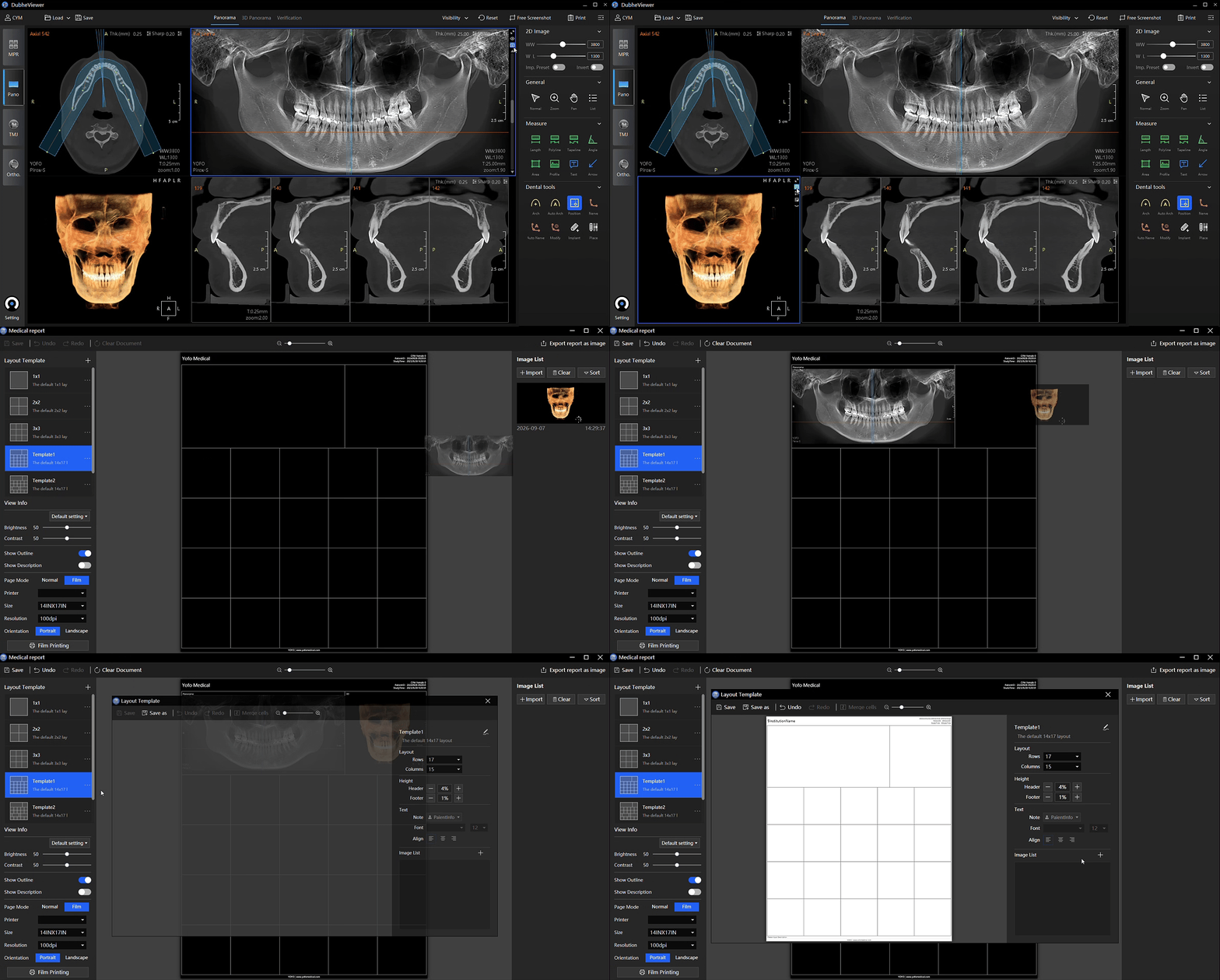
Custom Report Template Editor
Create professional, fully customizable imaging reports for panoramic, cephalometric and TMJ examinations. Customize layouts, clinic logos, typography and comments to create distinctive, branded reports that elevate your practice's professional brand.
Designed to simplify complexity. Built to elevate every decision.
_1.png) 40%R&D Team
40%R&D Team
 80%Proprietary
_1.png) 300+Patented CBCT
300+Patented CBCT 20+Countries Served
 1,000+CBCT units
 NO.1in market share for high-end CBCT





_1.png)